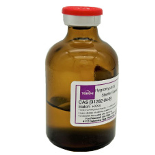
Hygromycin B, EvoPure solution (100 mg/mL in PBS Buffer) is formulated with highly pure (>99.0%) Hygromycin B. Hygromycin B is a unique aminoglycoside antibiotic derived from Streptomyces hygroscopicus and is routinely used as a selective agent in cell culture or microbiology applications to isolate Hygromycin B resistant cells after transfection or transformation, respectively.
This product is considered a dangerous good. Quantities above 1 g may be subject to additional shipping fees. Please contact us for questions.
For more Hygromycin B products, click here.
For more information on our EvoPure line of products, click here.
| Mechanism of Action |
Hygromycin B inhibits protein synthesis by strengthening the interaction of tRNA binding in the ribosomal A-site. Hygromycin B also prevents mRNA and tRNA translocation by an unknown mechanism. These are unique mechanisms for an aminoglycoside antibiotic and they differ from the mode of action of Neomycin, Gentamicin, and G418. Mechanism of Resistance Hygromycin B resistance is conferred by the hph gene isolated from Streptomyces hygroscopicus , a 1467 bp fragment which encodes Hygromycin B phosphotransferase (HPh). Cell lines successfully transfected with the hph gene produce hygromycin B phosphotransferase and convert hygromycin B to 7”-O-phosphoryl-hygromycin B by phosphorylating the 4-hydroxyl group on the cyclitol ring of hygromycin B. 7”-O-phosphoryl-hygromycin B lacks antibiotic activity and does not interact with prokaryotic or eukaryotic ribosomes. |
| Spectrum | Hygromycin B is effective against eukaryotic and prokaryotic cells. |
| Microbiology Applications | Hygromycin B can be used as a selection agent to isolate Hygromycin B resistant bacteria and fungi. The following Hygromycin B selection concentrations should serve as a guide only and may vary depending on experimental conditions and cells used: o Bacteria (E. coli) - 50 µg/mL - 100 µg/mL o Fungi - 100 µg/mL - 300 µg/mL o Yeasts - 50 µg/mL - 200 µg/mL |
| Plant Biology Applications | Hygromycin B is routinely used as a selection agent for Arabidopsis plants that have been transformed with a hygromycin B resistance gene. A rapid method to screen for hygromycin B resistant Arabidopsis in less than four days has been developed. After Arabidopsis seeds have been transformed with a resistance plasmid (pBIG-HYG), they are plated on MS medium with hygromycin B and subjected to a two day stratification at 4°C in the dark. Seeds are then exposed to light for 4-6 hours to stimulate germination and then placed in the dark for another two days. Transformed seeds are selected and identified after a 24 hour period in the light. Resistant transformants are characterized by long hypocotyls. (Harrison et al, 2006). |
| Eukaryotic Cell Culture Applications | Hygromycin B is routinely used as a selective agent in mammalian cell culture to isolate hygromycin B resistant cells after transfection. Selectable markers for hygromycin B resistant cells include the hyg or hph resistance genes which express a phosphotransferase that inactivates hygromycin B by phosphorylation. Effective working concentrations range from 100 – 1000 µg/mL.
For additional information regarding relevant cell lines, resistance plasmids, and culture media, please visit our cell culture database. |
| Molecular Formula | C20H37N3O13 |
| Documents | Hygromycin Protocol.pdf|Plasmid_DNA_Transfection_Protocol.pdf|Selection_of_Stable_Transfected_Cell_Lines_Protocol.pdf |
| References |
Dai S et al (2001) Comparative analysis of transgenic rice plants obtained by Agrobacterium-mediated transformation and particle bombardment. Mol. Breeding. 7: 25–33 González A, Jiménez A, Vázquez D, Davies JE, Schindler D. (1978) Studies on the mode of action of hygromycin B, an inhibitor of translocation in eukaryotes. Biochim Biophys Acta. 521(2):459-469 PMID 367435 |
| Protocols |
Hygromycin B Kill Curve ProtocolBackground: Hygromycin B is a unique aminoglycoside antibiotic produced by Streptomyces hygroscopicus and is routinely used as a selective agent in cell culture and microbiology applications to isolate transfected, hygromycin B resistant cells. Before stable transfected cell lines can be selected, the optimal hygromycin B concentration needs to be determined by performing a kill curve titration. The optimal working concentration of hygromycin B suitable for selection of resistant mammalian clones depends on the cell lines, media, growth conditions, and the quality of hygromycin B. Because of these variables, it is necessary to perform a kill curve for every new cell type and new batch of hygromycin B. Preparation and storage of hygromycin B solution: Hygromycin B is soluble in water at >50 mg/mL. It is also soluble in methanol or ethanol. Solutions should be sterilized by filtration, not by autoclaving. Hygromycin B solutions have been reported to lose activity on freezing. Since solutions are stable refrigerated, freezing should be avoided. Hygromycin B products should be stored as supplied at 2-8 °C. The dry solid is stable for at least five years if stored at 2-8 °C. Hygromycin B solutions are stable as supplied for two years if stored at 2-8 °C. Kill Curve/Hygromycin B Titration Protocol:
Plasmid DNA Transfection Protocol
Once the appropriate antibiotic concentration to use for selection of the stable transfected cells has been determined by performing a kill curve, the next step is to generate a stable cell line by transfection of the parental cell line with a plasmid containing the gene of interest and an antibiotic resistance gene.
Plasmid DNA Transfection Protocol:
QC Seed 24-wells with insert and determine the transfection efficiency by immunostaining:
Selection of Stable Transfected Cell Lines ProtocolBackground: Once the cells have been successfully transfected, the next step is to seed and select the transfected cell line in a single 96-well plate to select pure colonies by limited dilution as outlined below:
Protocol:
QC Seed 24-wells with insert for an immunostaining to determine percentage of cells expressing the gene of interest to be able to identify a 100% pure clone. You can also use Western blotting, flow cytometry or another technique depending on the cell line used. Seed 24-wells with insert and determine the expression level of the gene of interest by immunostaining:
|


