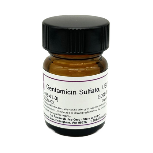
Gentamicin Sulfate, USP is an aminoglycoside antibiotic complex that was discovered in 1963 derived from fermentation of Micromonospora purpurea or M. echinospora. Gentamicin is composed of different components including Gentamicin C complex (Gentamicin C1, Gentamicin C1a, and Gentamicin C2) which makes up 80% of the compound and has the highest antibacterial activity, along with Gentamicin A, B, X, and a few others which make up the remaining 20% of Gentamicin and have lower antibiotic activity. Gentamicin Sulfate is suitable for use in cell culture to prevent and control bacterial contamination. Gentamicin, USP is is soluble in water.
Gentamicin Sulfate, USP conforms to United States Pharmacopoeia specifications.
The Centers for Disease Control (CDC) Standard Operating Procedure (SOP) DSR-052-04 includes the addition of Gentamicin Sulfate to viral transport media.
We also offer:
- Gentamicin Sulfate, EP (G007)
- Gentamicin Sulfate ReadyMadeTM Solution (G046)
- Gentamicin A Sulfate, EvoPure® (G035)
- Gentamicin C1 Sulfate, EvoPure® (G031)
- Gentamicin C1a Sulfate, EvoPure® (G032)
- Gentamicin C2 Sulfate, EvoPure® (G033)
- Gentamicin C2a Sulfate, EvoPure® (G034)
- Gentamicin C2b Sulfate, EvoPure® (G062)
- Gentamicin X2 Sulfate, EvoPure® (G036)
| Mechanism of Action | Aminoglycosides are a widespread and versatile group of bioactive natural products. They target the 30S ribosomal subunit, blocking the translocation of peptidyl-tRNA from acceptor to donor. This results in an inability to read mRNA ultimately producing a faulty or nonexistent protein. |
| Spectrum | Gentamicin Sulfate is a broad-spectrum antibiotic targeting Gram-positive and Gram-negative bacteria. It is effective against several strains of Mycoplasma. It also combats certain β-lactam sensitive VRE or vancomycin resistant Enterococcus; a "superbug." |
| Impurity Profile | Gentamicin C1: 25.0 -50.0% Gentamicin C1a: 10.0 - 35.0% Gentamicin C2a + C2: 25.0 - 55.0% |
| Microbiology Applications | Gentamicin Sulfate is commonly used as a selective agent to select for cells containing the Gentamicin resistance gene, aacj-AaphD or aacC1. Gentamicin Sulfate is generally used at a concentration of 10 - 50 µg/mL for eukaryotic cell culture and 15 ug/ml for prokaryotic cell culture.
According to the CDC SOP (DSR-052-04) for Viral Transport Medium (VTM), Gentamicin Sulfate is used at a final concentration of 100 µg/ml. Media SupplementsGentamicin can be used as a selective agent in several types of isolation media: Columbia Blood Agar - Gardnerella vaginalis Selective Supplement VRE Medium - VRE Selective Supplement Burkholderia cepacia Agar Base - Burkholderia cepacia Selective Supplement Microfluidic droplet systems are popular analytical methods used in antimicrobial susceptibility testing (AST) to help study antimicrobial resistance (AMR). Droplet methods have many advantages over conventional AST methods. Authors used several antibiotics from TOKU-E including Gentamicin Sulfate to review the physicochemical properties to predict retention of antibiotics in droplets (Ruszczak et al, 2023). |
| Plant Biology Applications | Gentamicin Sulfate inhibited differentiation of tracheary elements in pith parenchyma cells in cultures of romaine lettuce (Lactuca sativa L. var. Romana) at concentrations of 50-100 µg/ml. Similar results were obtained with cultured explants of Jerusalem artichoke tuber (Helianthus tuberosus L.). Callus formation was suppressed with increasing levels of Gentamicin Sulfate in both tissue systems. When studying cell division or xylem differentiation in culture, it is recommended to use ≤ 10 µg/ml according to published sources. |
| Eukaryotic Cell Culture Applications | Gentamicin was found to inhibit Glucose-6-phosphate dehydrogenase (G6PD) enzyme activity in rat erythrocytes (Temel et al, 2018). A bovine macrophage (Bomac) cell line was used but proved to be contaminated with BVDV (bovine viral diarrhea virus). Both infected and cured cells were tested for uptake of Mycoplasma bovis in an in vitro model to dissect the molecular and cellular details of bovine mycoplasmosis using Gentamicin (TOKU-E) in a gentamicin protection assay (Burgi et al, 2018). Gentamicin is an effective in vitro bacterial inhibitor that is nontoxic in tissue culture. It was non-toxic to RMK (Rhesus monkey kidney), HeLa, amnion, GMK, and WI-38 cell lines. It can also be used in virus tissue culture as it does not inhibit virus replication (Rudin et al, 1970). Porcine intestinal epithelial cell line IPEC-J2 cell monolayers were used to study the toxicity of Gentamicin in cell culture. Researchers found it did not affect IPEC-J2 cell monolayer integrity via the disruption of cell membranes and did not increase the incidence of toxic effects related to membrane permeation (Gyetvai et al, 2015). When primary cultures of embryonic rat fibroblasts are exposed to Gentamicin, they develop typical lysosomal phospholipidosis characterized by decreased sphingomyelinase activity, increase in lipid phosphorus, and appearance of 'myeloid bodies' in lysosomes. However, when inhibitors of cysteine proteinases were used (leupeptin, E-64), authors observed a protective effect which could be due to increased sphingomyelinase activity (Montenez et al, 1984). |
| Cancer Applications | Ovarian melanoma tumor cells was studied in 3D culture and Gentamicin Sulfate was used to prevent contamination when studying ovarian cell lines (OVCAR3, SKOV3, 222, EG, and A2780-PAR ) and normal ovarian surface epithelial cell lines (HIO 1120 and HIO 180). Tumor cells formed matrix-rich tubular networks containing channels surrounding spheroids of tumor cells, and this network may represent either a primitive microcirculatory-like network, or a remodeled vascularized portion of a tumor (Sood et al, 2001). |
| References |
Deng et al used Gentamicin Sulfate (TOKU-E) in Gentamicin protection assays to evaluate effect of miR-30c-1-3p and miR-30c-5p on intracellular Helicobacter pylori. Link to article. Bürgi N, Josi C, Bürki S and Schweizer, Pilo P (2018) Mycoplasma bovis co-infection with bovine viral diarrhea virus in bovine macrophages. Vet. Res. 49(1):2. PMID 29316971 Centers for Disease Control and Prevention. 2020. DSR-052-02: Preparation of viral transport medium. Link to SOP. Deng Q et al (2022) miR-30c increases the intracellular survival of Helicobacter pylori by inhibiting autophagy. Cellular Microbiol. 4536450 Gyetvai B et al (2015) Gentamicin Sulphate permeation through porcine intestinal epithelial cell monolayer. Act. Vet. Hung. 63(1): 60-68 PMID 25655415 Ruszczak A, Jankowski P, Vasantham S, Scheler O and Garstiecki P (2023) Physicochemical properties predict retention of antibiotics in water-in-oil droplets. Anal. Chem. 95(2): 1574−1581 PMID 36598882
|
| MIC | Bacillus cereus| 1.22|| Bacillus subtilis| 0.5|| Escherichia coli| 1.22 - 2.44 || Klebsiella pneumonia| 1|| Pseudomonas aeruginosa| 0.5 - 2.44 || Salmonella enterica| 1|| Staphylococcus aureus| 0.5 - 0.61 || Stenotrophomonas maltophilia| 2 - >16 || |


